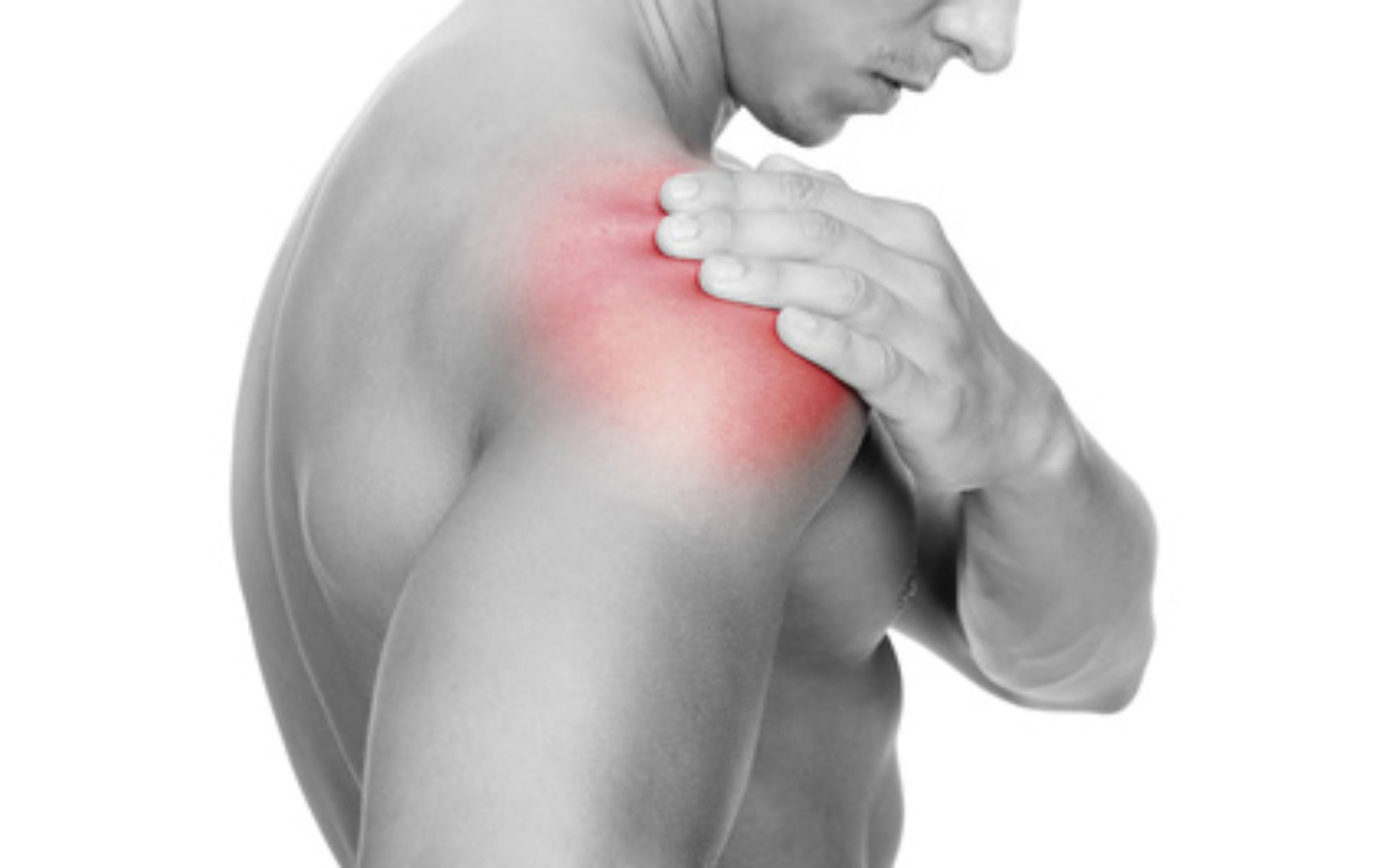
Hip pain is a common and disabling condition that affects patients of all ages. The differential diagnosis of hip pain is broad, presenting a diagnostic challenge. Patients often express that their hip pain is localized to one of three anatomic regions: the anterior hip and groin, the posterior hip and buttock, or the lateral hip. Anterior hip and groin pain is commonly associated with intra-articular pathology, such as osteoarthritis and hip labral tears. Posterior hip pain is associated with piriformis syndrome, sacroiliac joint dysfunction, lumbar radiculopathy, and less commonly ischiofemoral impingement and vascular claudication. Lateral hip pain occurs with greater trochanteric pain syndrome.
Clinical examination tests, although helpful, are not highly sensitive or specific for most diagnoses; however, a rational approach to the hip examination can be used. Radiography should be performed if acute fracture, dislocations, or stress fractures are suspected. Initial plain radiography of the hip should include an anteroposterior view of the pelvis and frog-leg lateral view of the symptomatic hip. Magnetic resonance imaging should be performed if the history and plain radiograph results are not diagnostic. Magnetic resonance imaging is valuable for the detection of occult traumatic fractures, stress fractures, and osteonecrosis of the femoral head. Magnetic resonance arthrography is the diagnostic test of choice for labral tears.
Hip pain is a common presentation in primary care and can affect patients of all ages. In one study, 14.3% of adults 60 years and older reported significant hip pain on most days over the previous six weeks.1 Hip pain often presents a diagnostic and therapeutic challenge. The differential diagnosis of hip pain (Table A) is broad, including both intra-articular and extra-articular pathology, and varies by age. A history and physical examination are essential to accurately diagnose the cause of hip pain.
Differential Diagnosis of Hip Pain
| DIAGNOSIS | PAIN CHARACTERISTICS | HISTORY/RISK FACTORS | EXAMINATION FINDINGS | ADDITIONAL TESTING |
|---|---|---|---|---|
|
Anterior thigh pain |
||||
|
Meralgia paresthetica |
Paresthesia, hypesthesia |
Obesity, pregnancy, tight pants or belt, conditions with increased intra-abdominal pressure |
Anterior thigh hypesthesia, dysesthesia |
None |
|
Anterior groin pain |
||||
|
Athletic pubalgia (sports hernia) |
Dull, diffuse pain radiating to inner thigh; pain with direct pressure, sneezing, sit-ups, kicking, Valsalva maneuver |
Soccer, rugby, football, hockey players |
No hernia, tenderness of the inguinal canal or pubic tubercle, adductor origin, pain with resisted sit-up or hip flexion |
Radiography: No bony involvement |
|
MRI: Can show tear or detachment of the rectus abdominis or adductor longus |
||||
|
Anterolateral hip and groin pain (C sign) |
||||
|
Femoral neck fracture/stress fracture |
Deep, referred pain; pain with weight bearing |
Females (especially with female athlete triad), endurance athletes, low aerobic fitness, steroid use, smokers |
Painful ROM, pain on palpation of greater trochanter |
Radiography: Cortical disruption |
|
MRI: Early bony edema |
||||
|
Femoroacetabular impingement |
Deep, referred pain; pain with standing after prolonged sitting |
Pain with getting in and out of a car |
FADIR and FABER tests are sensitive |
Radiography: Cam or pincer deformity, acetabular retroversion, coxa profunda |
|
Hip labral tear |
Dull or sharp, referred pain; pain with weight bearing |
Mechanical symptoms, such as catching or painful clicking; history of hip dislocation |
Trendelenburg or antalgic gait, loss of internal rotation, positive FADIR and FABER tests |
MRI: Can show a labral tear |
|
Magnetic resonance arthrography: offers added sensitivity and specificity |
||||
|
Iliopsoas bursitis (internal snapping hip) |
Deep, referred pain; intermittent catching, snapping, or popping |
Ballet dancers, runners |
Snap with FABER to extension, adduction, and internal rotation; reproduction of snapping with extension of hip from flexed position |
Radiography: No bony involvement |
|
MRI: Bursitis and edema of the iliotibial band |
||||
|
Ultrasonography: Tendinopathy, bursitis, fluid around tendon |
||||
|
Dynamic ultrasonography: Snapping of iliopsoas or iliotibial band over greater trochanter |
||||
|
Legg-Calvé-Perthes disease |
Deep, referred pain; pain with weight bearing |
2 to 12 years of age, male predominance |
Antalgic gait, limited ROM or stiffness |
Radiography: Early small femoral epiphysis, sclerosis and flattening of the femoral head |
|
Loose bodies and chondral lesions |
Deep, referred pain; painful clicking |
Mechanical symptoms, history of hip dislocation or low-energy trauma, history of Legg-Calvé-Perthes disease |
Limited ROM, catching and grinding with provocative maneuvers, positive FADIR and FABER tests |
Radiography: Can show ossified or osteochondral loose bodies |
|
MRI: Can detect chondral and fibrous loose bodies |
||||
|
Osteoarthritis of the hip |
Deep, aching pain and stiffness; pain with weight bearing |
Older than 50 years, pain with activity that is relieved with rest |
Internal rotation < 15 degrees, flexion < 115 degrees |
Radiography: Presence of osteophytes at the acetabular joint margin, asymmetrical joint-space narrowing, subchondral sclerosis and cyst formation |
|
Osteonecrosis of the hip |
Deep, referred pain; pain with weight bearing |
Adults: Lupus, sickle cell disease, human immunodeficiency virus infection, corticosteroid use, smoking, and alcohol use; insidious onset, but can be acute with history of trauma |
Pain on ambulation, positive log roll test, gradual limitation of ROM |
Radiography: Femoral head lucency and subchondral sclerosis, subchondral collapse (i.e., crescent sign), flattening of the femoral head |
|
MRI: Bony edema, subchondral collapse |
||||
|
Slipped capital femoral epiphysis |
Deep, referred pain; pain with weight bearing |
11 to 14 years of age, overweight (80th to 100th percentile) |
Antalgic gait with foot externally rotated on occasion, positive log roll and straight leg raise against resistance tests, pain with hip internal rotation relieved with external rotation |
Radiography: Widened epiphysis early, slippage of femur under epiphysis later |
|
Septic arthritis |
Refusal to bear weight, pain with leg movement |
Children: 3 to 8 years of age, fever, ill appearanceAdults: Older than 80 years, diabetes mellitus, rheumatoid arthritis, recent joint surgery, hip or knee prostheses |
Guarding against any ROM; pain with passive ROM |
Hip aspiration guided by fluoroscopy, computed tomography, or ultrasonography; Gram stain and culture of joint aspirate |
|
MRI: Useful for differentiating septic arthritis from transient synovitis |
||||
|
Transient synovitis |
Refusal to bear weight |
Children: 3 to 8 years of age, sometimes fever and ill appearance |
Pain with extremes of ROM |
|
|
Lateral pain |
||||
|
External snapping hip* |
Pain with direct pressure, radiation down lateral thigh, snapping or popping |
All age groups, audible snap with ambulation |
Positive Ober test, snap with Ober test, pain over greater trochanter |
Radiography: No bony involvement |
|
MRI: Bursitis and edema of the iliotibial band |
||||
|
Ultrasonography: Tendinopathy, bursitis, fluid around tendon |
||||
|
Greater trochanteric bursitis* |
Pain with direct pressure, radiation down lateral thigh |
Runners, middle-aged women |
Pain over greater trochanter |
Dynamic ultrasonography: Snapping of iliopsoas or iliotibial band over greater trochanter |
|
Greater trochanteric pain syndrome |
Pain with direct pressure, radiation down lateral thigh |
Associated with knee osteoarthritis, increased body mass index, low back pain; female predominance |
Proximal iliotibial band tenderness, Trendelenburg gait is sensitive and specific |
|
|
Posterolateral pain |
||||
|
Gluteal muscle tear or avulsion* |
Pain with direct pressure, radiation down lateral thigh and buttock |
Middle-aged women |
Weak hip abduction, pain with resisted external rotation, Trendelenburg gait is sensitive and specific |
MRI: Gluteal muscle edema or tears |
|
Iliac crest apophysis avulsion |
Tenderness to direct palpation |
History of direct trauma, skeletal immaturity (younger than 25 years) |
Iliac crest tenderness and/or ecchymosis |
Radiography: Apophysis widening, soft tissue swelling around iliac crest |
|
Posterior pain |
||||
|
Hamstring muscle strain or avulsion |
Buttock pain, pain with direct pressure |
Eccentric muscle contraction while hip flexed and leg extendedSkeletal immaturity, eccentric muscle contraction (cutting, kicking, jumping) |
Ischial tuberosity tenderness, ecchymosis, weakness to leg flexion, palpable gap in hamstring |
Radiography: Avulsion or strain of hamstring attachment to ischium |
|
Ischial apophysis avulsion |
Buttock pain, pain with direct pressure |
MRI: Hamstring edema and retraction |
||
|
Ischiofemoral impingement |
Buttock or back pain with posterior thigh radiation, sciatica symptoms |
Groin and/or buttock pain that may radiate distally |
None established |
MRI: Soft tissue edema around quadratus femoris muscle |
|
Piriformis syndrome |
Buttock pain with posterior thigh radiation, sciatica symptoms |
History of direct trauma to buttock or pain with sitting, weakness and numbness are rare compared with lumbar radicular symptoms |
Positive log roll test, tenderness over the sciatic notch |
MRI: Lumbar spine has no disk herniation, piriformis muscle atrophy or hypertrophy, edema surrounding the sciatic nerve |
|
Sacroiliac joint dysfunction |
Pain radiates to lumbar back, buttock, and groin |
Female predominance, common in pregnancy, history of minor trauma |
FABER test elicits posterior pain localized to the sacroiliac joint, sacroiliac joint line tenderness |
Radiography: Possibly no findings, narrowing and sclerotic changes of the sacroiliac joint space |
FABER = flexion, abduction, external rotation; FADIR = flexion, adduction, internal rotation; MRI = magnetic resonance imaging; ROM = range of motion.
*—Conditions associated with greater trochanteric pain syndrome.
Anatomy
The hip joint is a ball-and-socket synovial joint designed to allow multiaxial motion while transferring loads between the upper and lower body. The acetabular rim is lined by fibrocartilage (labrum), which adds depth and stability to the femoroacetabular joint. The articular surfaces are covered by hyaline cartilage that dissipates shear and compressive forces during load bearing and hip motion. The hip’s major innervating nerves originate in the lumbosacral region, which can make it difficult to distinguish between primary hip pain and radicular lumbar pain.
The hip joint’s wide range of motion is second only to that of the glenohumeral joint and is enabled by the large number of muscle groups that surround the hip. The flexor muscles include the iliopsoas, rectus femoris, pectineus, and sartorius muscles. The gluteus maximus and hamstring muscle groups allow for hip extension. Smaller muscles, such as gluteus medius and minimus, piriformis, obturator externus and internus, and quadratus femoris muscles, insert around the greater trochanter, allowing for abduction, adduction, and internal and external rotation.
In persons who are skeletally immature, there are several growth centers of the pelvis and femur where injuries can occur. Potential sites of apophyseal injury in the hip region include the ischium, anterior superior iliac spine, anterior inferior iliac spine, iliac crest, lesser trochanter, and greater trochanter. The apophysis of the superior iliac spine matures last and is susceptible to injury up to 25 years of age.2
Evaluation of Hip Pain
HISTORY
Age alone can narrow the differential diagnosis of hip pain. In prepubescent and adolescent patients, congenital malformations of the femoroacetabular joint, avulsion fractures, and apophyseal or epiphyseal injuries should be considered. In those who are skeletally mature, hip pain is often a result of musculotendinous strain, ligamentous sprain, contusion, or bursitis. In older adults, degenerative osteoarthritis and fractures should be considered first.
Patients with hip pain should be asked about antecedent trauma or inciting activity, factors that increase or decrease the pain, mechanism of injury, and time of onset. Questions related to hip function, such as the ease of getting in and out of a car, putting on shoes, running, walking, and going up and down stairs, can be helpful.3 Location of the pain is informative because hip pain often localizes to one of three basic anatomic regions: the anterior hip and groin, posterior hip and buttock, and lateral hip (eFigure A).

Figure A.
Localization of hip pain. (A) Posterior view. (B) Anterior view.
Taken from Am Fam Physician. 2014 Jan 1;89(1):27-34., https://www.aafp.org/afp/2014/0101/p27.html